Le Infezioni in Medicina, n. 3, 407-410, 2023
doi: 10.53854/liim-3103-16
CASE REPORTS
A case of histological diagnosis of Toxoplasma gondii myositis in a person living with HIV
Ilaria De Benedetto1, Francesco Biagini2, Guido Urbano3, Tiziana Enrica Mongini3, Ilaria Cassetta2, Luca Scaglione2, Antonio Curtoni4, Guido Calleri5, Andrea Calcagno1, Francesco Giuseppe De Rosa1,6, Silvia Corcione1,7
1Department of Medical Sciences, Infectious Diseases, University of Turin, Turin, Italy;
2Internal Medicine Unit, University Hospital Città della Salute e della Scienza di Torino, Turin, Italy;
3Neuromuscular Center, University Hospital Città della Salute e della Scienza di Torino, Department of Neurosciences RLM, University of Turin, Turin, Italy;
4Microbiology and Virology Unit, University Hospital Città della Salute e della Scienza di Torino, Turin, Italy;
5Department of Infectious Diseases, Travel Medicine Unit, Amedeo di Savoia Hospital, Torino, Italy;
6Infectious Diseases Unit, Cardinal Massaia Hospital, Asti, Italy;
7Tufts University School of Medicine, Boston, MA, USA
Article received 8 may 2023, accepted 15 August 2023
Corresponding author
Ilaria De Benedetto
E-mail: ilaria.debenedetto@unito.it
SummaRY
We report the case of a 58-year-old male with a recent diagnosis of HIV infection admitted for progressive muscular weakness and psychomotor impairment. Cerebrospinal examination documented a mild hyperproteinorrachia, with normal cells count and reduced glycorrhachia. Brain gadolinium-enhanced MRI showed bilateral T2 and FLAIR hyperintensities in the nucleo-capsular region and irregular contrast-enhancement of the globi pallidi and the right putamen. The histologic analysis of a quadriceps biopsy showed several foci of inflammatory infiltrates with concomitant muscular fiber atrophy and degeneration. Scattered intracytoplasmic inclusions were observed in muscle fibers, representing the main pathological feature. A positive PCR for Toxoplasma gondii and a Toxoplasma gondii specific monoclonal antibody immunohistochemical staining confirmed the diagnosis.
Keywords: HIV; Toxoplasma gondii; myositis; muscle; intracytoplasmic inclusion.
A 58-year-old male with a recent diagnosis of HIV infection (CD4+ 26 cells/mm³ after 4 months from ART initiation with tenofovir alafenamide, emtricitabine and dolutegravir), prophylaxis with Trimethoprim/Sulfamethoxazole three days a week and a previous diagnosis of Guillain-Barré syndrome treated with intravenous immunoglobulins was admitted for a 1-week-history of progressive weakness and mild psychomotor impairment associated with diarrhea. On admission, the patient was febrile (37.8°C), with normal blood pressure 110/70, peripheral oxygen saturation 94%, pulse rate 100/minute. On laboratory exams: CPK 2300 UI/Ln (n.v. <190), C-reactive Protein 10 mg/L (normal values <5), GOT 117 UI/L, GPT 45 UI/L, Troponin 85 ng/L and NT-proBNP 516 ng/L, normal blood count; on the lymphocyte subset analysis CD4+ 8 cells/mm³ (normal values 493-1666, <5%), CD4+/CD8+ ratio 0.1 (normal values 1.4-2.4); HIV-1 RNA 721 copies/mL. At neurological assessment, the patient presented severe symmetrical proximal weakness, inability to stand or sit independently, and initial dysphagia. Brain contrast CT scan showed a new hypodensity in the right nucleo-capsular region with mild contrast enhancement in the central part. A lumbar puncture was performed: CSF was clear and documented a mild hyperproteinorrachia (total proteins 95 mg/dl) with normal cells count (<2 elements/mmc), glycorrhachia was low, CSF culture was negative. T3 gadolinium-enhanced brain MRI showed T2 and FLAIR bilateral hyperintensities of the nucleo-capsular region - with right prevalence - and an irregular contrast-enhancement of the globi pallidi and the right putamen, without any increase in relative cerebral blood volume or restricted diffusion (Figure 1). Nerve conduction study documented a chronic axonal sensory-motor polyneuropathy. Treatment with intravenous immunoglobulins showed no benefit on the patient’s condition and the screening for myositis-specific and myositis-associated antibodies tested negative. A right quadriceps muscle biopsy was performed: the histological analysis showed several foci of inflammatory infiltrates - mainly composed of CD8 positive T-cells - with concomitant muscular fiber atrophy and degeneration. Scattered intracytoplasmic inclusions were observed in muscle fibers, representing the main pathological feature (Figure 2 and 3). The biopsy was performed to differentiate between a para-infectious myositis, a seronegative necrotizing autoimmune myopathy and an antiretroviral toxic myopathy. The histological examination of the muscle biopsy was compatible with Toxoplasma localization and a positive PCR for Toxoplasma gondii on muscle sample through Toxoplasma gondi ELITe MGB® Kit (ELITechGroup SAS, Puteaux, France) and a Toxoplasma gondii specific monoclonal antibody immunohistochemical staining confirmed the diagnosis (Figure 4). In detail, the tissue sample was frozen in isopentane cooled in liquid nitrogen, as for standard procedure. Routine histochemical staining were performed on 8-micron cryostat sections. Toxoplasma gondii-specific monoclonal antibodies (Virostat - Monotope, dilution 1:50) were applied to formalin-fixed muscle sections and incubate overnight. A secondary antibody conjugated to a DAB (diaminobenzidine)-based detection system was applied. The stained tissue sections were then examined under a light microscope.
Moreover, Cryptosporidium antigen stool test, serology for Strongyloides, blood culture tested negative. VDRL and viral PCR (including EBV and JCV DNA) tested negative on CSF while Toxoplasma gondii DNA was detected. The patient started the specific therapy against Toxoplasma with pyrimethamine and sulfadiazine, and a medium dosage intravenous corticosteroid therapy was then started (0.75 mg/kg/day), followed by oral maintenance with remarkable clinical results. An echocardiography did not disclose any cardiac involvement. Laboratory exams documented the normalization of CK (109 UI/L). At discharge, the patient was able to perform assisted exercises of the limbs and active trunk training exercises.
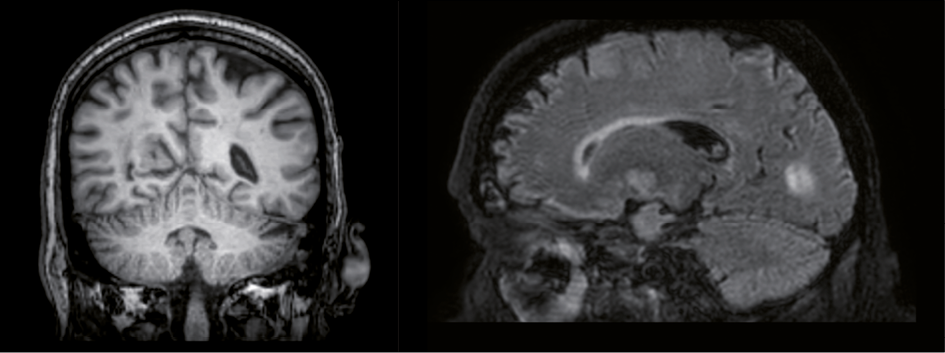
Figure 1 - T3 gadolinium-enhanced brain MRI showing Toxoplasma gondii lesions, T1 (left) and T2 (right).
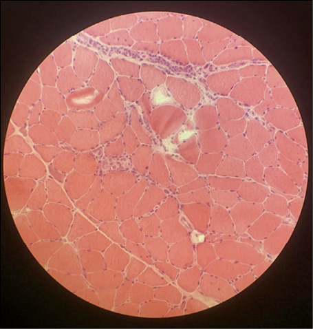
Figure 2 - Muscular biopsy in haematoxylin and eosin staining, 10x.
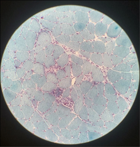
Figure 3 - Muscular biopsy in Gomori’s Trichrome.
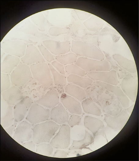
Figure 4 - Formalin-fixed muscle section stained with anti-Toxoplasma Gondii antibodies.
DISCUSSION
Toxoplasma gondii is a protozoan parasite that infects most species of animals, including humans. In the human host, the parasites form tissue cysts, most commonly in skeletal muscle, myocardium, brain, and eyes which may remain throughout the life of the host [1]. Following an acute infection, tachyzoites enter the brain and other tissues they are attacked by an immune response, nonetheless the parasite can persist as intraneuronal or intramuscular cyst-forming bradyzoites which occasionally reactivate depending on the host’s immune status, possibly causing encephalitis or myositis [2]. Bradyzoites are difficult to be addressed by drugs that cannot penetrate the interior of the tissue cyst and they avoid immune-mediated destruction [3]. Clinical manifestations of muscular localization are rare even in the case of immune-compromised hosts and only few cases have been reported in the last 30 years [4-10]. In 1992, five cases were described in patients with HIV with marked lymphopenia with less than five CD4+ cells/mm3 [4]. Another case of histologically confirmed Toxoplasma myositis in a non-HIV-infected patient was reported in 2003 in a patient with idiopathic CD4 lymphocytopenia [5]. Other two cases were described in Brazilian siblings: one developed a tetraplegia that was confirmed to be due to inflammatory myositis due to toxoplasma, the other developed myocarditis, with heart failure, without skeletal muscle weakness; in both cases Toxoplasma organisms were observed in the muscle biopsies [6]. To date no effective treatment is reported to eradicate bradyzoites and Toxoplasma myositis should be included among differentials of sudden muscular weakness, especially in immune-compromised host. In this case, a possible role for immunoglobulins treatment of Guillame-Barré syndrome in reactivation of Toxoplasma cannot be ruled out. In this contest of limited therapeutic approaches, prevention of acquisition, screening and possibly primary prophylaxis should primarily be addressed in key populations.
Conflict of interest
Authors have no conflict of interests related to disclose.
Funding
This research was supported by EU funding within the MUR PNRR Extended Partnership initiative on Emerging Infectious Diseases (Project no. PE00000007, INF-ACT)
Author contributions
I.D.B. and F.G. wrote the paper; I.D.B, G.U., T.E.M., A.C., A.C., S.C. and F.G.D.R. revised the paper; I.C., L.S. and T.E.M. had the patient in charge; F.G.D.R, S.C and L.S. supervised the work; S.C. coordinated the work.
Ethical conduct of research statement
Permission to anonymous publication has been obtained through signed consent by the patient.
REFERENCES
- Centers for Disease Control and Prevention (CDC)- https://www.cdc.gov/parasites/toxoplasmosis/biology.html
- Wohlfert EA, Blader IJ, Wilson EH. Brains and Brawn: Toxoplasma infections of the central nervous system and skeletal muscle. Trends Parasitol. 2017; 33 (7), 519-531. doi: 10.1016/j.pt.2017.04.001.
- Tomita T, et al. Making Home Sweet and Sturdy: Toxoplasma gondii ppGalNAc-Ts glycosylate in hierarchical order and confer cyst wall rigidity. MBio. 2017; 8 (1).
- Gherardi R, Baudrimont M, Lionnet F, et al. Skeletal muscle toxoplasmosis in patients with acquired immunodeficiency syndrome: a clinical and pathological study. Ann Neurol. 1992; 32 (4), 535-542. doi: 10.1002/ana.410320409.
- Plonquet A, Bassez G, Authier FJ, Dray JM, Farcet JP, Gherardi RK. Toxoplasmic myositis as a presenting manifestation of idiopathic CD4 lymphocytopenia. Muscle Nerve. 2003; 27 (6), 761-765. doi: 10.1002/mus.10376.
- Calore EE, Minkovski R, Khoury Z, et al. Skeletal muscle pathology in 2 siblings infected with Toxoplasma gondii. J Rheumatol. 2000; 27 (6), 1556-1559. PMID: 10852291.
- Montoya JG, Jordan R, Lingamneni S, et al. Toxoplasmic myocarditis and polymyositis in patients with acute acquired toxoplasmosis diagnosed during life. Clin Infect Dis. 1997; 24, 676-683.
- Hassene A, Vital A, Anghel A, et al. Acute acquired toxoplasmosis presenting as polymyositis and chorioretinitis in immunocompetent patient. Joint Bone Spine. 2008; 75, 603-605.
- Cuomo, G., D’Abrosca, V., Rizzo, V. et al. Severe polymyositis due to Toxoplasma gondii in an adult immunocompetent patient: a case report and review of the literature. Infection. 2013; 41, 859-862. https://doi.org/10.1007/s15010-013-0427-x
- Leroy J, Houzé S, Dardé ML, et al. Severe toxoplasmosis imported from tropical Africa in immunocompetent patients: A case series. Travel Med Infect Dis. 2020; 35, 101509. doi: 10.1016/j.tmaid.2019.101509.